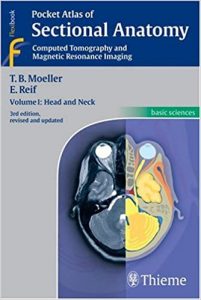
Renowned for its superb illustrations and highly practical information, the third edition of this classic reference reflects the very latest in state-of-the-art imaging technology. Together with Volumes 2 and 3, this compact and portable book provides a highly specialized navigational tool for clinicians seeking to master the ability to recognize anatomical structures and accurately interpret CT and MR images.
Features:
- New CT and MR images of the highest quality
- Didactic organization using two-page units, with radiographs on one page and full-color illustrations on the next
- Concise, easy-to-read labeling on all figures
- Color-coded, schematic diagrams that indicate the level of each section
- Sectional enlargements for detailed classification of the anatomical structure
Comprehensive, compact, and portable, this book is ideal for use in both the classroom and clinical setting.
Volume II: Thorax, Heart, Abdomen, and Pelvis
